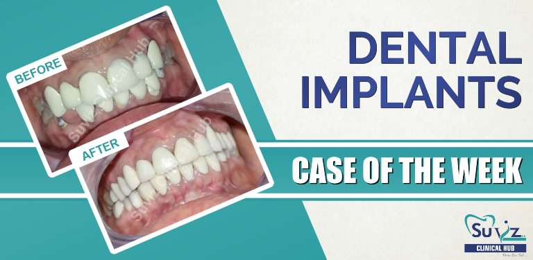
Age: 42 Gender: Female
Type of Case: Full mouth implant rehabilitation
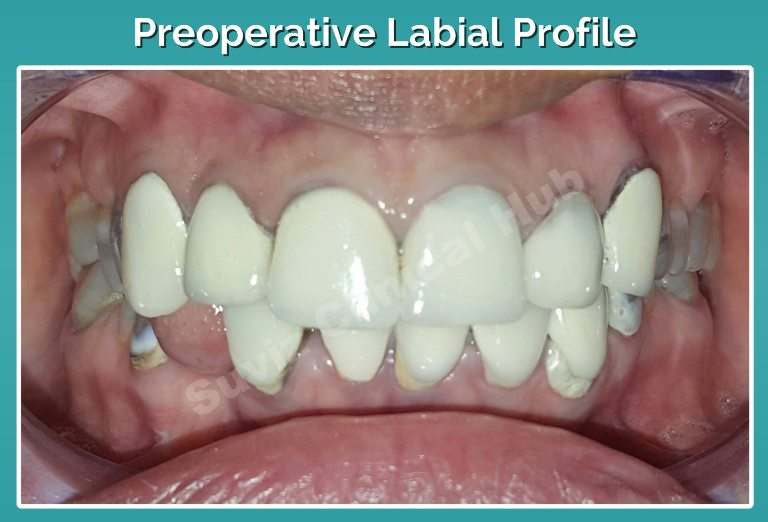
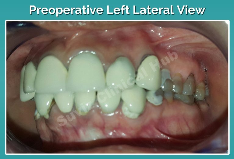
Chief Complaint: Metal show of the caps on upper front teeth and missing lower front tooth
Dental History: Upper and lower anterior PFM crowns were fabricated 10 yrs ago.
Pre Operative:
Clinical Findings:
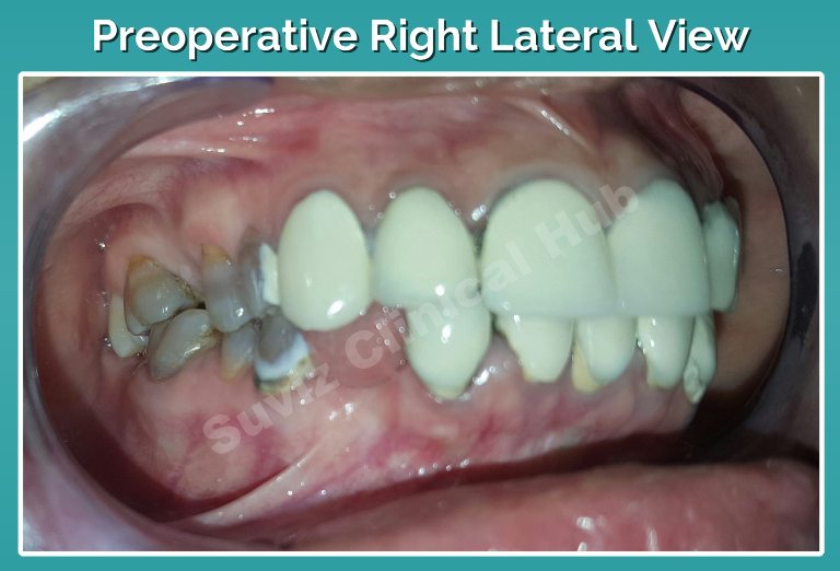
- Existing PFM crowns wrt upper and lower anteriors.
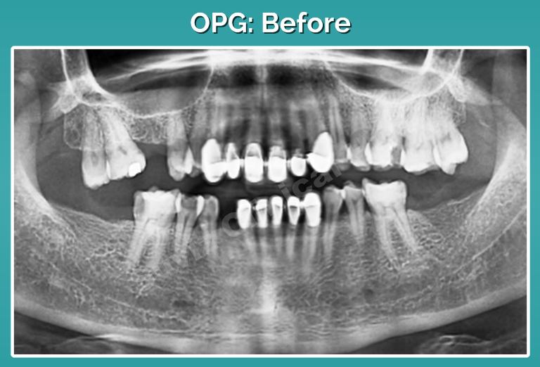
- Missing 16,37,38,43,47,48
- Deep dental caries wrt 26, 46.
During The Procedure:
Alternative Treatment Plan:
- Replacement of the upper and lower anterior crowns using Emax crowns.
- Replacement of missing teeth by FPD or an RPD.
Treatment Plan:
- Root canal treatment wrt 26, 46
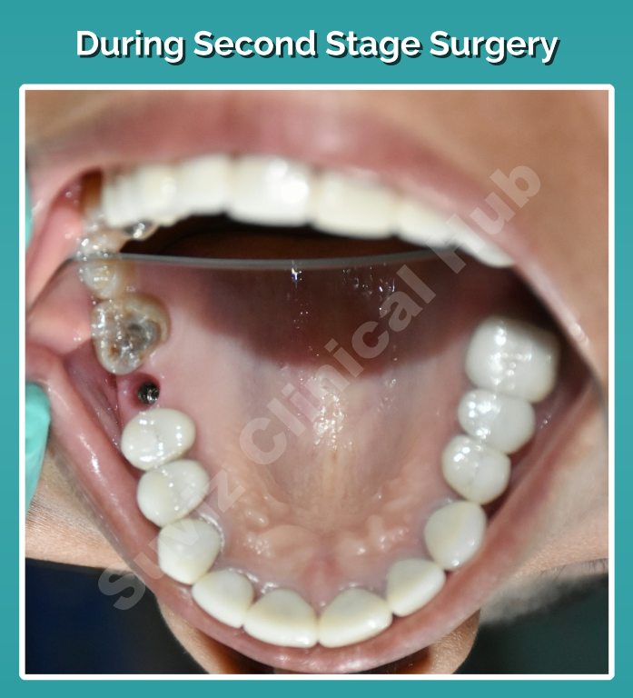
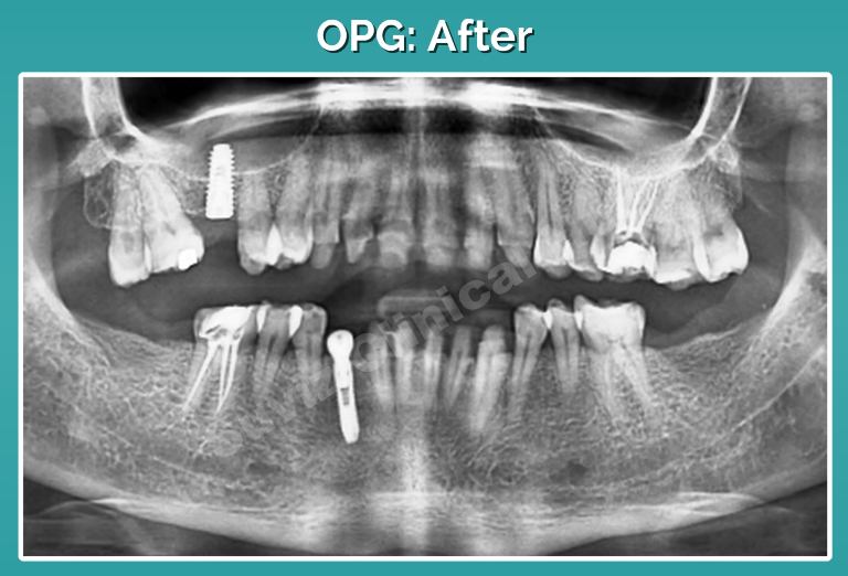
- Surgical Implant placement wrt 16,43.
- Extraction wrt 17,18,27,28.
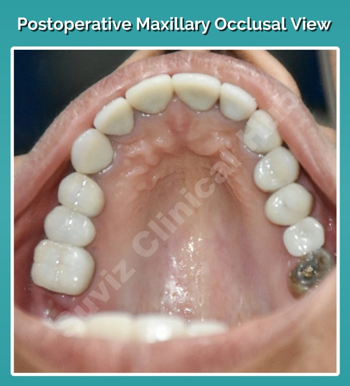
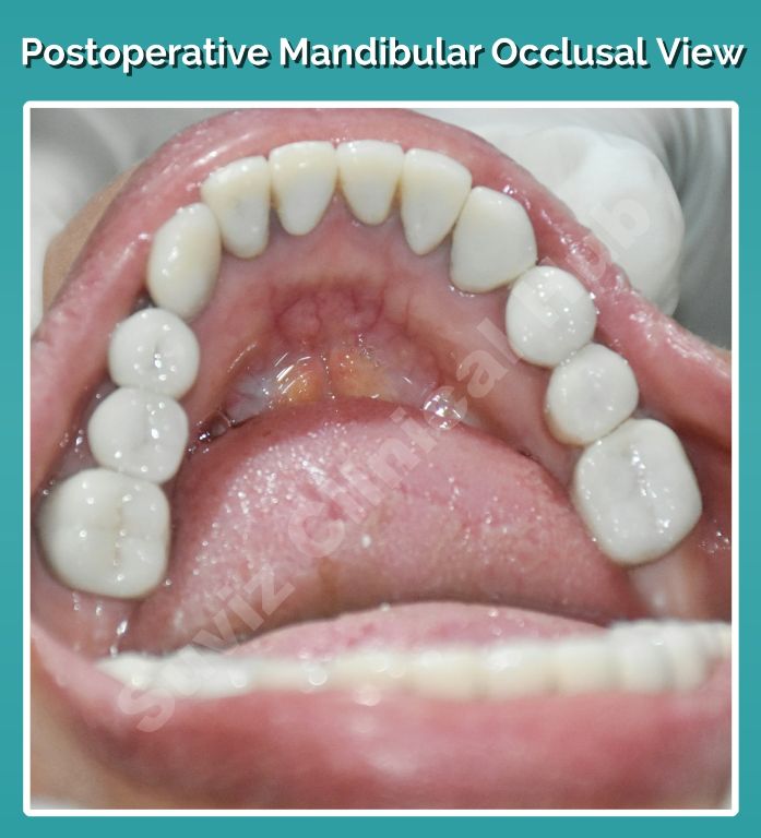
- Full mouth rehabilitation using full veneer crowns and implant restoration.
Treatment Procedure:
- Preoperative models and photographs were taken.
- The face-bow transfer was made, centric, protrusive and lateral occlusal records were obtained.
- The casts were mounted onto a semi-adjustable articulator and programmed. Broadricks analysis was made to establish the occlusal plane.
- A mock-up was performed.
- Clinically, surgical implant placement was done wrt 16 and 43.
- Sutures were removed in a week.
- The existing PFM crowns were sectioned and removed.
- They were temporized and anterior guidance was established.
- The lower posteriors were prepared and temporized and the occlusal plane was established.
- Following which, the upper posteriors were prepared and temporized.
- A mutually protected occlusion was achieved.
- They were kept under observation for 6 weeks before making the final impressions and the interocclusal records.
- The bisque trial of the all-ceramic crowns were done and satisfactory.
- The final cementation was carried out on the natural teeth using resin cement.
- The implant restorations were planned for a progressive loading by cement-retained crowns at the end of 2 months.
Post Operative:
Follow Up:
- Advised for a regular 6 months follow up.
